Gartner s duct cyst histology images are ready. Gartner s duct cyst histology are a topic that is being searched for and liked by netizens now. You can Download the Gartner s duct cyst histology files here. Get all royalty-free images.
If you’re looking for gartner s duct cyst histology images information connected with to the gartner s duct cyst histology topic, you have pay a visit to the right site. Our site always gives you suggestions for refferencing the highest quality video and picture content, please kindly surf and locate more informative video articles and graphics that fit your interests.
Although vaginal cysts are found in approximately 1 to 2 of women and gartner s duct cysts comprise approximately 10 of vaginal benign cysts 1 7 there is still some controversy regarding which course of action should be taken. She denies any pain or symptoms associated with the cyst. A gartner s duct cyst sometimes incorrectly referred to as vaginal inclusion cyst is a benign vaginal cyst that originates from the gartner s duct which is a vestigial remnant of the mesonephric duct wolffian duct in females. Like other cysts they are lined with non mucinous cuboidal or columnar epithelium. Vaginal inclusion cyst epidermal inclusion cyst squamous epithelium.
Gartner S Duct Cyst Histology. Bartholin s cyst squamous or columnar cells usu. A gartner s duct cyst sometimes incorrectly referred to as vaginal inclusion cyst is a benign vaginal cyst that originates from the gartner s duct which is a vestigial remnant of the mesonephric duct wolffian duct in females. The cyst type which was more frequently associated with symptoms was bartholin s duct cyst. Mullerian cysts were lined by columnar endocervical like or cuboidal epithelium whereas gartner s duct cysts were all lined by cuboidal epithelium.
 Figure 4 From Complex Malformations Of The Urogenital Tract In A Female Dog Gartner Duct Cyst Ipsilateral Renal Agenesis And Ipsilateral Hydrometra Semantic Scholar From semanticscholar.org
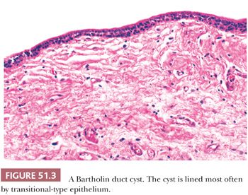
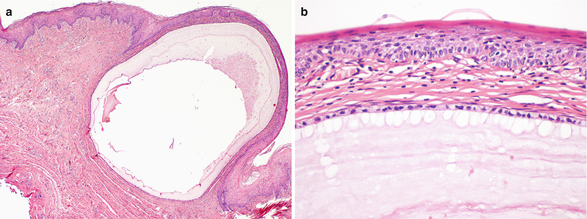
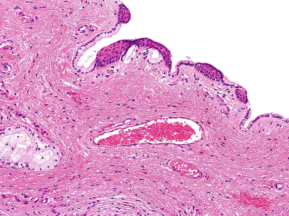
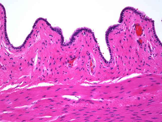
Aka epidermal inclusion cyst. They are typically small asymptomatic cysts that occur along the lateral walls of the vagina following the course of the duct. Histologic examination may be employed to correctly identify the cellular remnants composed of non mucin secreting low columnar or cuboidal epithelium but in clinical practice it is not necessary. Although vaginal cysts are found in approximately 1 to 2 of women and gartner s duct cysts comprise approximately 10 of vaginal benign cysts 1 7 there is still some controversy regarding which course of action should be taken. Mullerian cysts were lined by columnar endocervical like or cuboidal epithelium whereas gartner s duct cysts were all lined by cuboidal epithelium. Gartner duct cysts of vagina are types of vaginal cyst.
It is a benign cyst that is lined by non secretory cuboidal columnar epithelial cells when observed under a microscope by a pathologist.
Although vaginal cysts are found in approximately 1 to 2 of women and gartner s duct cysts comprise approximately 10 of vaginal benign cysts 1 7 there is still some controversy regarding which course of action should be taken. This is the first study reporting long term clinical observation of these lesions. During a routine pelvic exam a 20 year old woman has a 1 cm cystic mass incidentally discovered on the lateral wall of the vagina. Like other cysts they are lined with non mucinous cuboidal or columnar epithelium. She has no past medical history. Although vaginal cysts are found in approximately 1 to 2 of women and gartner s duct cysts comprise approximately 10 of vaginal benign cysts 1 7 there is still some controversy regarding which course of action should be taken.
 Source: basicmedicalkey.com
She denies any pain or symptoms associated with the cyst. It is a benign cyst that is lined by non secretory cuboidal columnar epithelial cells when observed under a microscope by a pathologist. Aka epidermal inclusion cyst. During a routine pelvic exam a 20 year old woman has a 1 cm cystic mass incidentally discovered on the lateral wall of the vagina. Like other cysts they are lined with non mucinous cuboidal or columnar epithelium.
 Source: twitter.com
During a routine pelvic exam a 20 year old woman has a 1 cm cystic mass incidentally discovered on the lateral wall of the vagina. Müllerian cyst endocervical epithelium. The cyst type which was more frequently associated with symptoms was bartholin s duct cyst. Gartner duct cysts of vagina are types of vaginal cyst. Aka epidermal inclusion cyst.
 Source: commons.wikimedia.org
Gartner duct cysts are located in the anterolateral wall of the proximal superior portion of the vagina 2 and are typically located above the level of the most inferior aspect of the pubic symphysis. A gartner s duct cyst sometimes incorrectly referred to as vaginal inclusion cyst is a benign vaginal cyst that originates from the gartner s duct which is a vestigial remnant of the mesonephric duct wolffian duct in females. Although vaginal cysts are found in approximately 1 to 2 of women and gartner s duct cysts comprise approximately 10 of vaginal benign cysts 1 7 there is still some controversy regarding which course of action should be taken. She denies any pain or symptoms associated with the cyst. They are typically small asymptomatic cysts that occur along the lateral walls of the vagina following the course of the duct.
 Source: commons.wikimedia.org
During a routine pelvic exam a 20 year old woman has a 1 cm cystic mass incidentally discovered on the lateral wall of the vagina. The cyst type which was more frequently associated with symptoms was bartholin s duct cyst. Like other cysts they are lined with non mucinous cuboidal or columnar epithelium. Gartner duct cysts of vagina are types of vaginal cyst. Vaginal inclusion cyst epidermal inclusion cyst squamous epithelium.
 Source: researchgate.net
Bartholin s cyst squamous or columnar cells usu. Aka epidermal inclusion cyst. The histology confirmed benign columnar epithelium consistent with a benign epithelial cyst and the diagnosis of a gartner s duct cyst was made. To define the course of the gartner s duct cyst and differentiate it from other pathologic considerations and structures mri can be a useful tool. Gartner duct cysts are located in the anterolateral wall of the proximal superior portion of the vagina 2 and are typically located above the level of the most inferior aspect of the pubic symphysis.
 Source: vetsci.org
Like other cysts they are lined with non mucinous cuboidal or columnar epithelium. These are also known as mesonephric cysts of vagina. Vaginal inclusion cyst epidermal inclusion cyst squamous epithelium. Like other cysts they are lined with non mucinous cuboidal or columnar epithelium. To define the course of the gartner s duct cyst and differentiate it from other pathologic considerations and structures mri can be a useful tool.
 Source: link.springer.com
A gartner s duct cyst sometimes incorrectly referred to as vaginal inclusion cyst is a benign vaginal cyst that originates from the gartner s duct which is a vestigial remnant of the mesonephric duct wolffian duct in females. Gartner duct cysts are located in the anterolateral wall of the proximal superior portion of the vagina 2 and are typically located above the level of the most inferior aspect of the pubic symphysis. She has no past medical history. This is the first study reporting long term clinical observation of these lesions. During a routine pelvic exam a 20 year old woman has a 1 cm cystic mass incidentally discovered on the lateral wall of the vagina.
 Source: link.springer.com
She has no past medical history. She has no past medical history. A gartner s duct cyst sometimes incorrectly referred to as vaginal inclusion cyst is a benign vaginal cyst that originates from the gartner s duct which is a vestigial remnant of the mesonephric duct wolffian duct in females. Most lesions were located in the left lateral vaginal wall 13 cases 32 50. Most common vaginal cyst.
 Source: sciencedirect.com
It is a benign cyst that is lined by non secretory cuboidal columnar epithelial cells when observed under a microscope by a pathologist. Gartner duct cysts of vagina are types of vaginal cyst. The histology confirmed benign columnar epithelium consistent with a benign epithelial cyst and the diagnosis of a gartner s duct cyst was made. The patient made an uneventful recovery and was symptom free at her check up 6 weeks post operatively. To define the course of the gartner s duct cyst and differentiate it from other pathologic considerations and structures mri can be a useful tool.
 Source: commons.wikimedia.org
She has no past medical history. A gartner s duct cyst sometimes incorrectly referred to as vaginal inclusion cyst is a benign vaginal cyst that originates from the gartner s duct which is a vestigial remnant of the mesonephric duct wolffian duct in females. It is a benign cyst that is lined by non secretory cuboidal columnar epithelial cells when observed under a microscope by a pathologist. She has no past medical history. Gartner duct cysts are located in the anterolateral wall of the proximal superior portion of the vagina 2 and are typically located above the level of the most inferior aspect of the pubic symphysis.
 Source: webpathology.com
The histology confirmed benign columnar epithelium consistent with a benign epithelial cyst and the diagnosis of a gartner s duct cyst was made. The histology confirmed benign columnar epithelium consistent with a benign epithelial cyst and the diagnosis of a gartner s duct cyst was made. This is the first study reporting long term clinical observation of these lesions. These are also known as mesonephric cysts of vagina. They are typically small asymptomatic cysts that occur along the lateral walls of the vagina following the course of the duct.
This site is an open community for users to do submittion their favorite wallpapers on the internet, all images or pictures in this website are for personal wallpaper use only, it is stricly prohibited to use this wallpaper for commercial purposes, if you are the author and find this image is shared without your permission, please kindly raise a DMCA report to Us.
If you find this site value, please support us by sharing this posts to your favorite social media accounts like Facebook, Instagram and so on or you can also bookmark this blog page with the title gartner s duct cyst histology by using Ctrl + D for devices a laptop with a Windows operating system or Command + D for laptops with an Apple operating system. If you use a smartphone, you can also use the drawer menu of the browser you are using. Whether it’s a Windows, Mac, iOS or Android operating system, you will still be able to bookmark this website.


